


 7 / 16

7
ESSM
Today
Key from KOLS: The Modified Sliding Technique (MOST) for penile length
and girth restoration in patients with severe erectile dysfunction
6. Ralph, D.J., et al.,
The immediate inser-
tion of a penile prosthesis for acute
ischaemic priapism
.
Eur Urol, 2009.
56(6): p. 1033-8
.
7. Wang, R., et al.,
Prospective and long-
term evaluation of erect penile length
obtained with inflatable penile pros-
thesis to that induced by intracaver-
nosal injection
.
Asian J Androl, 2009.
11(4): p. 411-5
.
8. Smith, J.F., et al.,
Risk factors for emo-
tional and relationship problems in
Peyronie’s disease
.
J Sex Med, 2008.
5(9): p. 2179-84
.
9. Egydio, P.H., F.E. Kuehhas, and S. Sansa-
lone,
Penile length and girth restora-
tion in severe Peyronie’s disease us-
ing circular and longitudinal grafting
.
BJU Int, 2013. 111(4 Pt B): p. E213-9
.
10. Sansalone, S., et al.,
Simultaneous
penile lengthening and penile pros-
thesis implantation in patients with
Peyronie’s disease, refractory erectile
dysfunction, and severe penile short-
ening.
J Sex Med, 2012. 9(1): p. 316-21
.
11. Rolle, L., et al.,
A new, innovative,
lengthening surgical procedure for
Peyronie’s disease by penile prosthe-
sis implantation with double dorsal-
ventral patch graft: the “sliding tech-
nique”
.
J Sex Med, 2012. 9(9): p. 2389-95
.
12. Egydio PH, Kuehhas FE.
Penile length-
ening and widening without graft-
ing according to a modified sliding
technique.
BJU Int. 2015 Jan 28. doi:
10.1111/bju.13065. [Epub ahead of print]
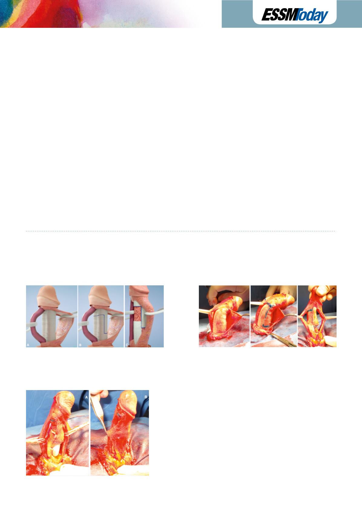
Fig. 1
Illustration of the preparation of the modified sliding technique:
A – Mobilization of the urethra and the neurovascular bundle through two longitudinal paraurethral incisions on Buck’s fascia
B – Marking of the “sliding edges”
C – The penis is stretched to its maximum length, limited only by the elasticity of the neurovascular bundle and the urethra
Fig. 2
A – Insertion of a penile prosthesis;
B – Coverage of the tunical defect with Buck’s fascia only.